Figure 3.
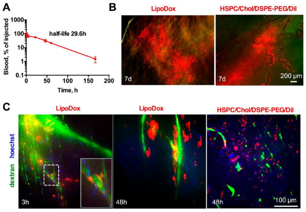
LipoDox (PEGylated liposomal doxorubicin) and PEGylated HSPC/Chol liposomes shows dynamics of deposition similar to PEGylated EPC liposomes. LipoDox was injected at 2 mg/kg (doxorubicin) into Balb/C mice. (A) Circulation half-life (monoexponential curve fit) of LipoDox in plasma (n = 2). (B) Low magnification ex vivo images of skin of mice injected with LipoDox or HSPC/Chol/DSPE-PEG-2000/DiI (7 days postinjection) show massive deposition of fluorescence. Some fluorescence follows the outside contour of blood vessels and the distribution of the liposomes is heterogeneous. (C) Intravital microscopy of skin of mice injected with LipoDox or HSPC/Chol/DSPE-PEG-2000/DiI shows localization in blood vessels and extravasation. The doxorubicin fluorescence, which was dimmer than DiI due to mismatch of Dox fluorescence with the optical setup of the intravital microscope, was enhanced in both images to the same extent. Animated 3-D images are provided as video 6 and video 7.

