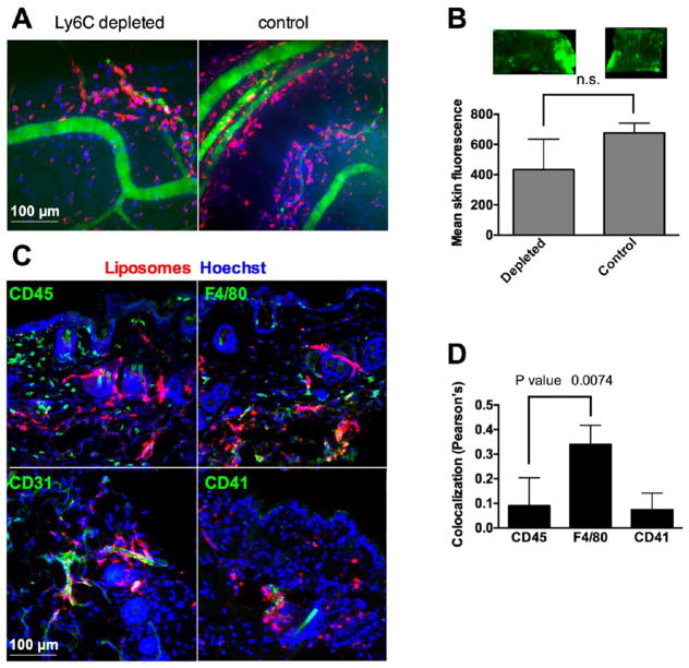
Figure 5.
Accumulation of PEGylated liposomes is independent of leukocytes and platelets. (A) Neutrophils were depleted with Ly6C-specific antibody as described in the Methods. Intravital imaging of skin 7 days postinjection shows similar extravasation and deposition of liposomes in depleted and control mice. (B) Representative scan of skin and mean fluorescence quantification show nonsignificant decrease in the accumulation in the depleted mice (p value 0.06, two-tailed unpaired t test, n = 3). (C) Immunostaining of skin histological sections 7 days postinjection shows minimal colocalization of liposomes with CD45+ leukocytes, CD41+ platelets, but colocalization with F4/80+ phagocytes (likely dendritic cells). Note that leukocytes are homogeneously distributed, F4/80+ cells are segregated in dermal and subdermal layers, and platelets are mostly limited to large blood vessels. Some liposomes were still located in the blood vessels (CD31+). (D) Colocalization analysis of the three stains in (C) with the liposomes (Pearson’s correlation coefficient r) using Coloc 2 plugin (ImageJ). F4/80 showed significantly more colocalization than CD45 (p = value 0.0074, nonpaired t test, 5–10 microscopical areas were analyzed).

