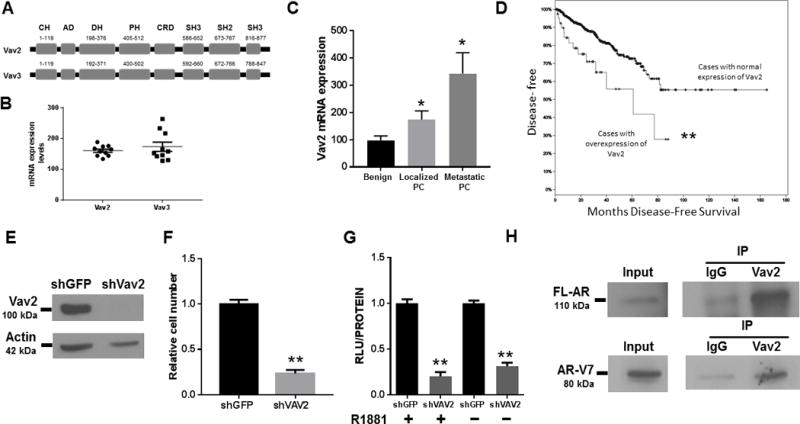
Figure 4. Expression of Vav2, a member of the Vav family, is elevated in human CRPC samples, and Vav2 interacts endogenously with and promotes the transcriptional activities of full length FL-AR and AR-V7.

A) A schematic of Vav2 and Vav3 structure with the amino acid position of each domain. CH = calponin homology domain, AD = acidic domain, DH = Diffuse B-Cell lymphoma homology (GEF) domain, PH = Pleckstrin homology domain, CRD = cysteine rich domain, SH2 = Src homology 2 domain, SH3 = Src homology 3 domain. B) Vav3 and Vav2 mRNA levels are co-expressed in human CRPC bone metastases which contain relatively high AR-V7 levels (dataset of Hornberg et al., 2011 (50)). C) Vav2 mRNA levels are elevated in PC and metastatic PC compared to benign tissue (dataset of Varambally et al., 2005). Kruskal Wallis test was performed, p = 0.02. D) Vav2 overexpression is prognostic for decreased disease-free survival. The Kaplan-Meier curve was built using the TCGA Prostate Adenocarcinoma dataset (n= 499). The upper curve denotes cases with no abnormal expression of Vav2, while the lower curve represents the cases in which Vav2 mRNA levels are upregulated (z-score threshold ± 2.0). P-value = 0.001. E) Immunoblotting was performed on 22Rv1 cell lysate using an anti-VAV2 antibody and anti-actin as the loading control. Data shown represent 1 of 2 independent experiments. F) Stable Vav2 depletion in 22Rv1 cells (vs control shGFP) decreased cell number. Data shown represent 2 independent experiments performed in quintuplicate. Independent Student’s T-test, p value < 0.001. G) 22Rv1 cells were transfected with the dual plasmid luciferase reporter system: MMTV or ΔGRE described in Materials and Methods. Luciferase activity was determined 48 h after transfection. Data shown represent 3 independent experiments performed in triplicate, showing the mean ± SE, and normalized to their shGFP controls. Unpaired T-test, p value = 0.001 in the presence of androgen; and, p value < 0.001 in the absence of androgen. H) 22Rv1 cells were harvested and co-immunoprecipitations were performed using antibodies to rabbit IgG control or Vav2. Immunocomplexes were immunoblotted with antibodies against FL-AR or AR-V7 (** p value < 0.01, * p value < 0.05).
