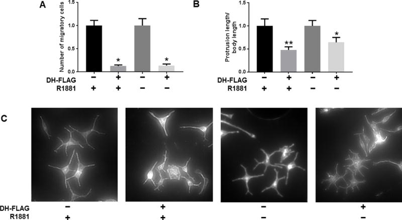
Figure 7. Disruption of coactivator interactions with FL-AR or AR-V7 decreases cell migration and introduces morphological changes.

A) 22Rv1 cells stably expressing DH-FLAG or empty vector linked to FLAG were seeded in Boyden Chambers for migration assays. Cells expressing DH-FLAG were normalized to their corresponding EV controls. Data show one experiment out of two performed in triplicate, showing the mean ± SE. For conditions with R1881: Independent T-test, p value = 0.01; for conditions without R1881: Independent T-test, p value = 0.02. B) The CRPC cell line C4-2B transiently expressing DH-FLAG or empty vector linked to were kept in vehicle or 1nM of R1881, and stained for Phalloidin immunofluorescence and DAPI. The total length of protrusions for each cell was measured and divided by cell body length. The average ± SE is shown. Cells expressing DH-FLAG were normalized to their EV controls. For conditions with R1881: n= 13, Mann Whitney test, p value = 0.008; for conditions without R1881: n = 15, Mann Whitney test, p value = 0.02. C) Representative images of C4-2B at 10X stained with Phalloidin. (** p value < 0.01, * p value < 0.05).
