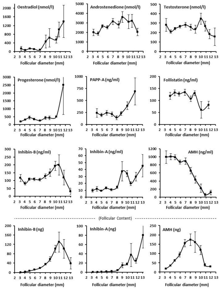
Figure 2.
Intrafollicular content and concentrations of peptide and steroid hormones. Graphs show the follicular fluid concentrations of estradiol, progesterone, androstenedione, testosterone, inhibin B, inhibin A, AMH, PAPP-A, and follistatin, and follicular content in nanograms (bottom panel) of inhibin B, inhibin A, and AMH according to follicular diameter. Values are presented as mean ± SEM. Statistically significant differences were observed in intrafollicular hormone levels of estradiol [analysis of variance (ANOVA); N = 264; P < 0.001], androstenedione (ANOVA; N = 276; P < 0.03), progesterone (ANOVA; N = 267; P < 0.003), PAPPA (ANOVA; N = 96; P < 0.03), inhibin-B (ANOVA; N = 627; P < 0.002), inhibin-A (ANOVA; N = 258; P < 0.001), AMH (ANOVA; N = 655; P < 0.001), but not testosterone (ANOVA; N = 566; P > 0.10) and follistatin (ANOVA; N = 148; P > 0.10). Regarding follicular content (bottom panel), the intrafollicular contents of inhibin-B (ANOVA; N = 611; P < 0.001), inhibin-A (ANOVA; N = 258; P < 0.001), and AMH (ANOVA; N = 654; P < 0.001) were observed to be significantly different in relation to follicular diameter. Specific numbers for the analyzed follicle groups divided according to follicular diameter are shown in Tables S1 and S2 in Supplementary Material.

