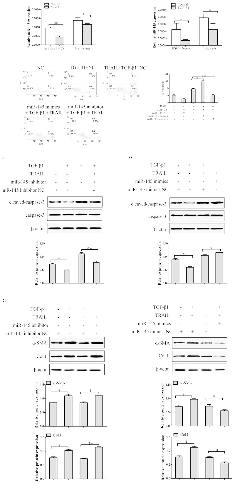
FIGURE 3.

The effect of miR-145 on the apoptosis of TGF-β1-treated LX-2 cells induced by TRAIL. (A) The expression of miR-145 was detected by quantitative real-time PCR in liver fibrosis tissues, primary HSCs and activated HSCs. (B) miR-145 inhibitor and miR-145 mimics, respectively decreased and increased the rate of apoptosis in activated LX-2 cells that were pretreated with TRAIL (10 ng/ml) for 24 h. (C) The protein expression of cleaved of caspase-3 was measured by Western blot in activated LX-2 cells transfected with miR-145 mimics that were pretreated with TRAIL (10 ng/ml) for 24 h. (D) The protein expression of cleaved of caspase-3 was assessed by Western blot in activated LX-2 cells transfected with miR-145 inhibitor that were pretreated with TRAIL (10 ng/ml) for 24 h. (E,F) The expression of α-SMA and Col. I protein was determined by Western blot in activated LX-2 cells that were pretreated with TRAIL (10 ng/ml) for 24 h. The assays were performed at least three times with similar results. Data are shown as the mean ± SD (n = 3) of one representative experiment. ∗p < 0.05, ∗∗p < 0.01 versus normal groups.
