Figure 5.
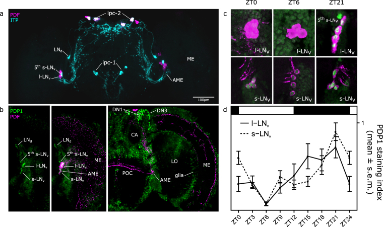
The clock network of B. oleae. (a) The clock neuropeptide PDF (magenta) is expressed in 4 l-LNv and 4 s-LNv, as well as in 4 putative insulin producing cells (ipc-2) in the PL. ITP (cyan) is expressed in the 5th s-LNv and in one cell of the LNd group, as well as in putative ipc-1 and ipc-2. (b) anti-PDP1 (green) co-localize in the nuclei of PDF positive cells (magenta) in the LNv cluster. Antibody reveals also other putative clock clusters (LNd, DN), and stains many other non-clock cells. (c,d) PDP1 protein level in the nuclei oscillates under LD12:12 in the s-LNv (H(7) = 34.78, p < 0.001) and l-LNv (H(7) = 19.259, p < 0.05). AME: accessory medulla; ME: medulla; CA: calyx; LO: lobula.

