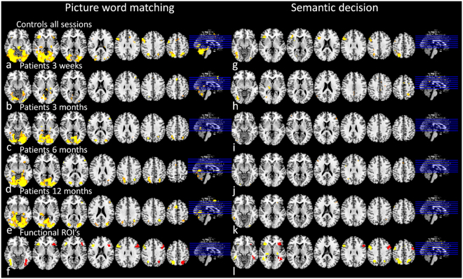
Figure 2.
Brain activation during language tasks. Average brain activation pattern in response to language tasks (p < 0.001, uncorrected; radiological orientation; left side is right hemisphere). For picture-word matching: in controls averaged over all sessions (a), in patients at session 1 (b), 2 (c), 3 (d) and 4 (e); and ROIs for subsequent analyses (f: original clusters (yellow), flipped to right hemisphere (red)). For semantic decision: in controls averaged over all sessions (g), in patients at session 1 (b), 2 (c), 3 (d) and 4 (e); and ROIs for subsequent analyses (f: original clusters (yellow), and areas flipped to right hemisphere (red)). IFG Inferior Frontal Gyrus, R right, L left, Bil bilateral, MNI Montreal Neurological Institute coordinates (X, Y and Z), t t-statistic of peak voxel within cluster.

