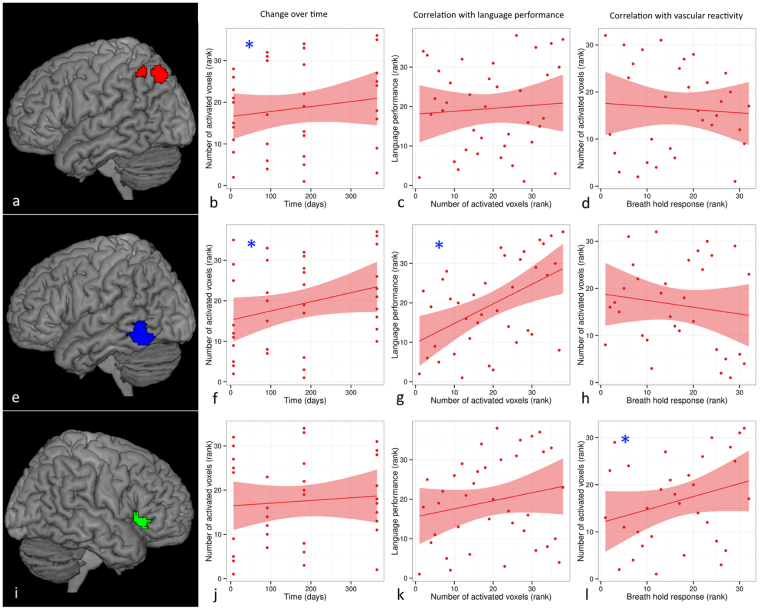
Figure 4.
Example of change of language activation over time in relation to language performance and vascular reactivity in stroke patients. Significant change of language activation (induced by a picture-word matching task) over time was found in left angular gyrus (red overlay (a)) (b) and left posterior inferior temporal gyrus (blue overlay (e)) (f), but not in right inferior frontal gyrus pars triangularis (green overlay (i)) (j). Language performance was significantly correlated with language activation in left posterior inferior temporal gyrus (g), but not in left angular gyrus (c) or right inferior frontal gyrus pars triangularis (k). Language activation was significantly correlated with vascular responsiveness (measured with a breath hold paradigm) in right inferior frontal gyrus pars triangularis (i), but not in left angular gyrus (d) or left posterior inferior temporal gyrus (h). See also Table 3. Statistical significance is indicated by [*].

