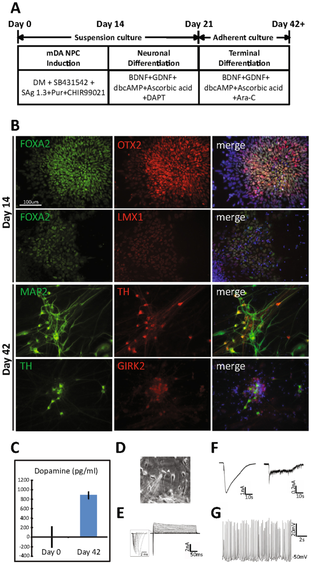
Figure 1.
mDA differentiation protocol yields mDA NPCs at day 14 and mDA neurons at day 42. (A) mDA differentiation scheme. After dissociation, iPS cells were kept in suspension culture for 21 days. In the first 14 days, cells were induced with DM (Dorsomorphin), SB431542, SAg 1.3 (Smoothened agonist), Pur (Purmorphamine), and CHIR99021. From day 14 through day 21, cells were differentiated in the neuronal differentiation medium containing BDNF, GDNF, dbcAMP, Ascorbic acid, and DAPT. From day 21, cells were further differentiated in the terminal differentiation medium containing BDNF, GDNF, dbcAMP, Ascorbic acid, and Ara-C. (B) Immunostaining of day 14 (top two rows) and day 42 (bottom two rows) 18a cells. (C) The mean concentration (pg/ml) of dopamine released by day 0 cells and day 42 18a cells. (D) Phase contrast image showing human iPSC 18a-derived dopaminergic neuron cultures after 1 month adherent culture. Arrowhead points to a recorded cell. (E) Representative traces showing whole-cell voltage-gated Na+ and K+ currents recorded in human iPSC 18a-derived dopaminergic neuron culture. (F) Representative traces showing responses to GABA and AMPA (100 representative traces each) (G) Representative traces showing spontaneous action potentials. The resting membrane potential was −50 mV.

