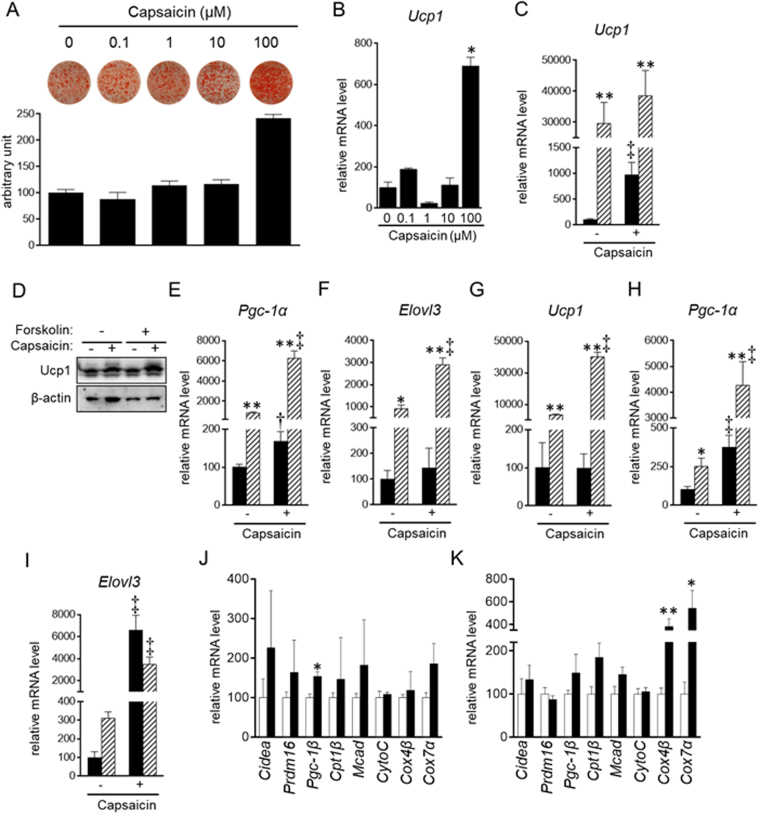
Figure 1.
Stimulation of brown adipogenesis by supra-pharmacological capsaicin. (A–F,J) HB2 brown preadipocytes or (G–I,K) SV cells from mouse brown adipose tissues were cultured with the indicated concentrations (A and B) or 100 μM (C–K) of capsaicin during brown adipogenesis. Cells on day 8 (C–F) or day 10 (G-I) were also treated with or without forskolin (10 μM) for 4 h. (A) Oil Red O staining of cells on day 8 was performed and the dye intensity was quantified (n = 2). Expression levels of Ucp1 (B,C,G), Pgc-1α (E and H) or Elolv3 (F and I) were examined by RT-qPCR analysis. (C,E–I) Black bar: vehicle; Hatched bar: forskolin. (J and K) Expression levels of genes related to brown adipogenesis and function of brown adipocytes were examined in HB2 brown adipocytes on day 8 (J) and primary mouse brown adipocytes on day 10 (K) in the absence of forskolin by RT-qPCR. Open bar: vehicle; Black bar: capsaicin. The data are presented as the mean ± SE (n = 4). *P < 0.05 and **P < 0.01 vs. cells treated with vehicle and corresponding reagent (vehicle or capsaicin). †P < 0.05 and ‡P < 0.01 vs. cells treated with vehicle and corresponding reagent (vehicle or forskolin). (D) Ucp1 as well as β-actin as the loading control was examined by Western blot analysis. Representative results are shown. The cropped images of Western blot analysis are shown because of space limitations; images of the full-length blot are Supplementary Fig. S6.

