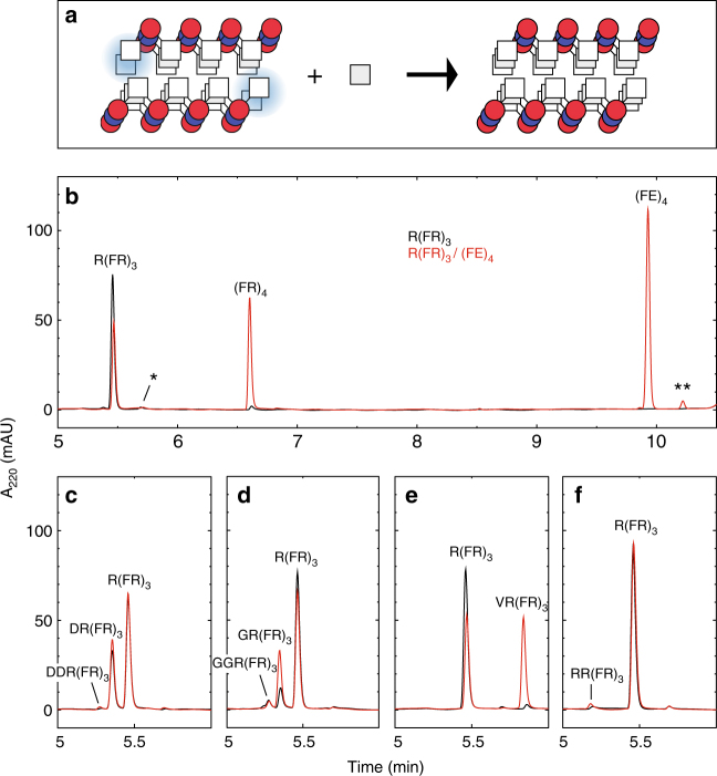
Fig. 1.
Single addition reactions with the R(FR)3/(FE)4 amyloid. a Schematic model of the amyloid-templated addition reaction, depicting just three layers of the repetitive amyloid structure in which the phenylalanine is represented by squares and the arginine and aspartate by blue and red circles respectively. The blue shading highlights the sites of addition in the substrate peptide. b–f, Reverse-phase HPLC chromatograms of the addition reactions with phenylalanine (b), aspartate (c), glycine (d), valine (e) and arginine (f). The * and ** indicate the C-terminal deamidated substrate and template, respectively, that remained after purification. The reactions with just the substrate R(FR)3 are the black traces and those with the amyloid R(FR)3/(FE)4 are the red ones. The R(FR)3 concentration was 100 μM, the (FE)4 concentration was 130 μM and amino acids were all at 100 μM

