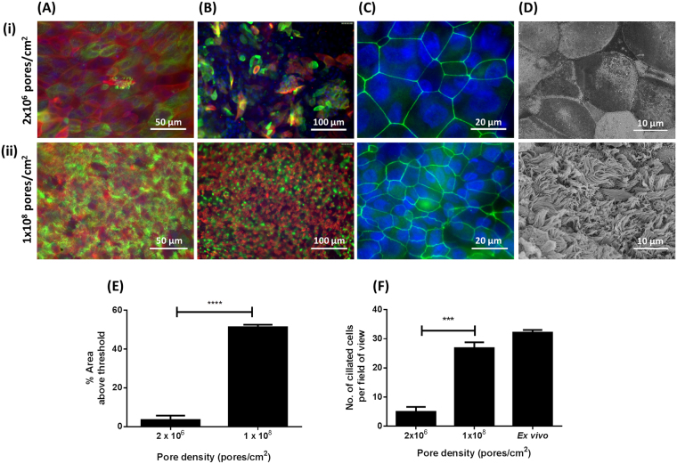
Figure 7.
Effect of cell culture insert pore density on cell differentiation. BBEC cultures were grown for 21 days at an ALI on membranes with pore densities of 2.0 × 10⁶ or 1.0 × 10⁸ pores/cm2 before fixation. The BBEC cultures were subsequently immunostained to assess (A) ciliation (cilia - green; F-actin - red; nuclei - blue), (B) mucus production (mucus - green; cilia - red; nuclei - blue) and (C) tight-junction formation (tight-junctions - green; nuclei - blue) or (D) examined by SEM. Representative images are shown of BBECs grown on inserts with (i) 2.0 × 10⁶ and (ii) 1.0 × 10⁸ pores/cm2. Quantitative analysis of ciliation of the apical surface of BBEC cultures was performed using (E) fluorescence intensity thresholding of immunostained cultures and (F) by counting the number of ciliated cells per field of view in H&E-stained sections as described in Fig. 2. Statistical significance was tested using an Ordinary one-way ANOVA: *** = P < 0.001; **** = P < 0.0001.

