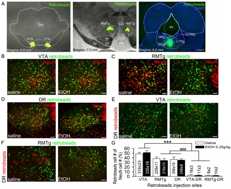
Figure 2.

LHb neurons project to the ventral tegmental area (VTA), rostral medial tegmental nucleus (RMTg), and dorsal raphe (DR). Images showing green retrobeads injected into the VTA (A, left panel), RMTg (A, middle panel) or DR (A, right panel) of ethanol naive rats. Ten days after injection, rats were randomly divided into two groups; one group received an intraperitoneal injection (i.p.) of 0.25 g/kg ethanol (EtOH), the other of saline (1 ml/Kg). Then the brain tissue was harvested for double staining of retrobeads and NeuN (a neuronal marker) to assess the distribution of retrobeads in the LHb. Intra-VTA retrobead injection resulted in a strong retrograde labeling in the LHb (B). Intra-RMTg or DR injection of retrobeads resulted in a moderate labeling in the LHb (C and D). Intra-VTA and DR or RMTg and DR dual-retrobeads injections resulted in a minority of LHb neurons labeled with green and red retrobeads (E and F). Compared with saline group, acute ethanol (0.25 g/kg, i.p.) did not significantly alter the ratios of neurons projecting to these areas. (G) Summary graph of percentages of cell counting from 5 sections of each rat (n=5/group). The percentage was calculated by (retrobeads labeling cells/NeuN cells) ×100%. Cell numbers are indicated in the bars. ***p < 0.001 vs. RMTg, DR, VTA-DR or RMTg-DR; ###p < 0.001 vs. VTA-DR or RMTg-DR, Bonferroni t-test followed by two-way ANOVA. 4v, 4th ventricle; Aq, aqueduct; CLi, the caudal linear nucleus of the raphe; DRc, dorsal raphe center; DTgP dorsal tegmental nucleus, pericentral part; IPN, interpeduncular nucleus; ml, medial lemniscus; PMnR, Paramedian raphe nucleus; tth, trigeminothalamic tract; VLPAG, ventrolateral periaqueductal gray. Scale bar =1 mm (A left panel-C right panel) and 20 μm (B-F).
