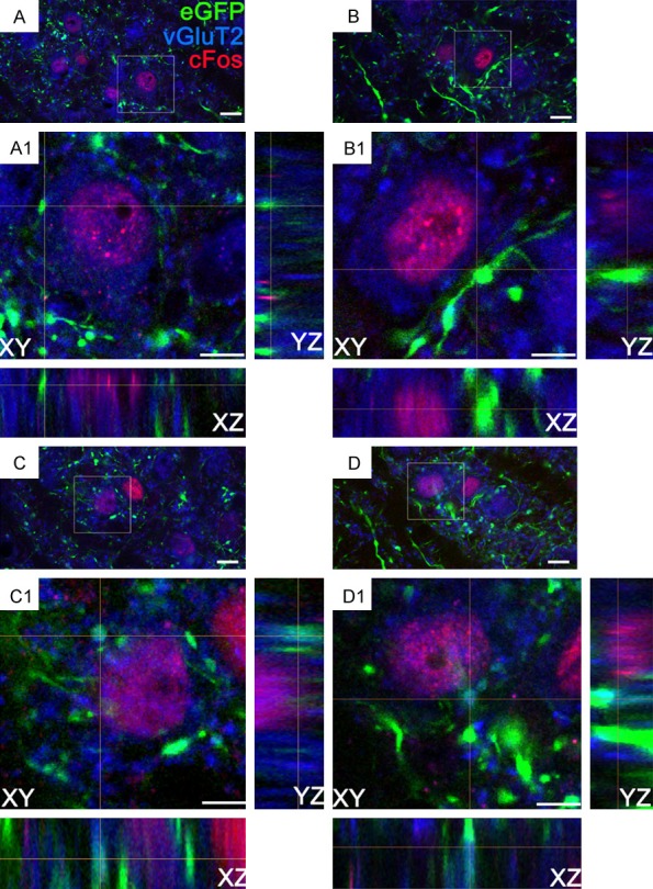
Figure 5.

Three-dimensional confocal images of LH axonal terminations in LHb vGluT2 cells expressing ethanol-induced cFos from rats that received an intra-LH injection of AAV5-CaMKIIa-eGFP, and ethanol (0.25 g/kg, i.p.) 2-3 weeks later. (A-D), show the LHb vGluT2+ cells (blue) expressing cFos (red), were contacted by axonal buttons (green) from the LH. Square boxes indicate the locations of the contacts. (A1-D1) show enlarged xy-, yz-, and xz- orthogonal views. Notably, (C, D) show the LH axonal varicosities expressing vGluT2 and, forming contact on LHb cFos+ cells. Since the confocal laser-scanning microscope generates in-focus images of selected depth, projection phenomena, in the conventional fluorescence microscope can be ruled out. Scale bars =10 μm (A-D) and 5 μm (A1-D1).
