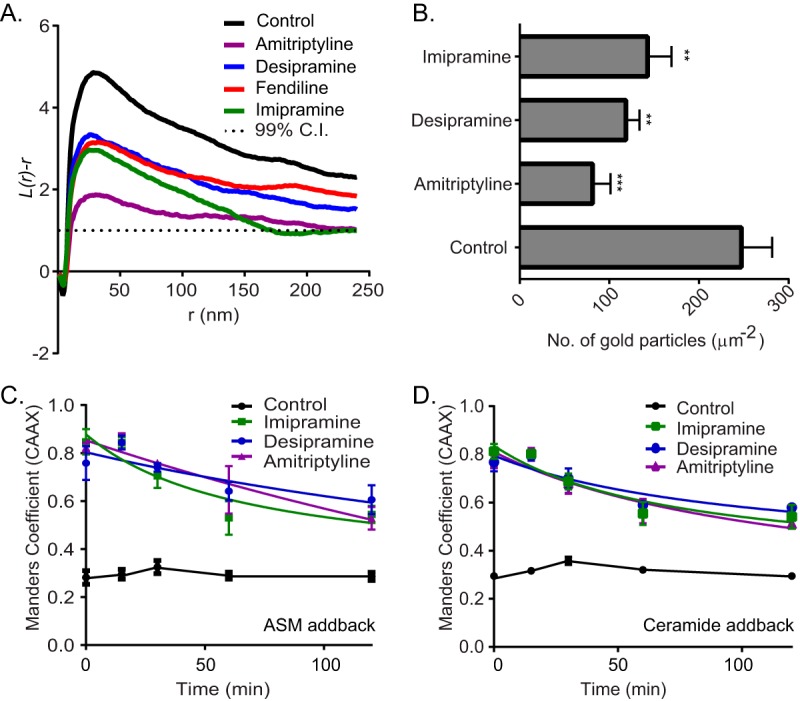
FIG 2.

ASM inhibitors reduce PM binding and nanoclustering of K-RasG12V. (A) Basal PM sheets prepared from MDCK cells expressing mGFP–K-RasG12V after treatment with vehicle (DMSO), imipramine (10 μM), desipramine (10 μM), or amitriptyline (1 μM) for 24 h were labeled with anti-GFP-conjugated gold particles. The spatial distribution of the gold particles visualized by EM was analyzed using univariate K functions. Plots of the weighted mean standardized univariate K functions are shown. (B) The graph shows mean numbers of gold particles in PM sheets ± standard errors of the means (n = 20). Significant differences were assessed by using Student's t tests (**, P < 0.01; ***, P < 0.001). (C and D) MDCK cells stably coexpressing mGFP–K-RasG12V and mCherry-CAAX were treated with vehicle (DMSO), imipramine (10 μM), desipramine (10 μM), or amitriptyline (1 μM) for 48 h and then incubated with recombinant ASM or 10 μM Cer in the continued presence of drugs and imaged by confocal microscopy. Cells were fixed at various time points, and PM localization of K-RasG12V was quantified by using Manders coefficients after imaging with a confocal microscope.
