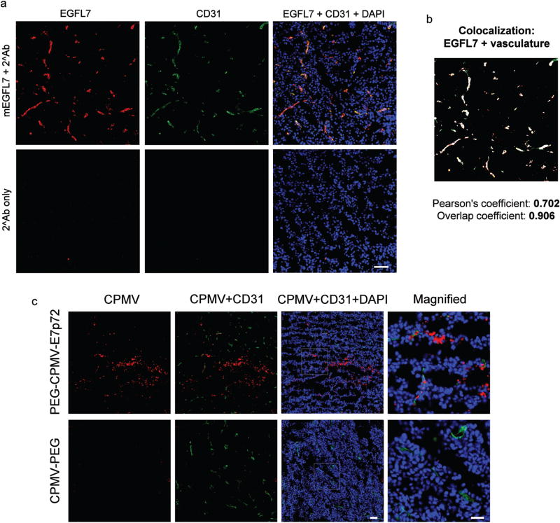
Fig. 6.
CPMV-PEG-E7p72 nanoparticle binds HT1080 tumor and neovasculature ex vivo. (a) Fluorescence images of HT1080 tumor tissues sections showing the expression of EGFL7 in tumor neovasculature. Immunofluorescence staining was performed to detect mouse EGFL7 (red), CD31 vascular marker (green), and nuclei (blue). Scale bar, 50 microns. (b) Composite image showing areas of mEGFL7 and CD31 localization (white). This image was generated using ImageJ (Colocalization Finder plugin). (c) Fluorescence images showing that CPMV-PEG-E7p72, but not CPMV-PEG nanoparticles bind to the tumor-associated neovasculature as well as HT1080 tumor tissues ex vivo. CPMV (red), CD31 (green), and nuclei (blue). Scale bar, 50 microns. Right panel shows high magnification of inset. Scale bar, 25 microns.

