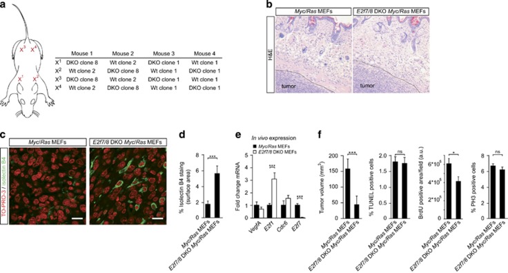
Figure 2.
Absence of E2F7/8 in mouse xenograft tumors leads to increased tumor vascularization.(a) Schematic representation of xenograft positions and experimental setup. MEFs were injected with a total of one million cells subcutaneously into athymic nude mice. The diameter of the tumors was monitored every 2 days. After 8 days, when the first tumors reached a diameter of 1 cm, all mice were euthanized and tumors harvested. (b) Hematoxylin and eosin (H&E) staining of control and E2f7/8 DKO xenograft tumors. Dashed black line indicates tumor border. (c) Staining and quantification (d) of intratumoral endothelial cell (Isolectin B4) and nuclei (TO-PRO-3) of control and E2f7/8 DKO xenograft tumors (n=8 tumors per genotype). For each tumor, five fields (× 400 magnification) were quantified. White scale bars in (c) indicate 10 μM. (e) Messenger RNA expression of Vegfa, E2f1, Cdc6 and E2f7 in harvested E2f7/8 DKO and control xenografted MEF tumors (n=8 tumors per genotype). Quantitative PCR analysis was used to analyze mRNA levels. Messenger RNA levels in E2f7/8 DKO tumors are depicted compared with control tumors. (f) Quantification (similar as in (d)) of tumor volume, in vivo BrdU incorporation, and phospho-histone 3 (PH3)-positive and TUNEL-positive cells as determined in the outer border zone of control and E2f7/8 DKO xenograft tumors. All quantified data present the average±s.e.m. compared with the indicated controls. *P<0,05; ***P<0,005; ns, not significant; a.u., arbitrary units.

