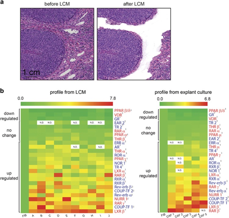
Figure 1.
NRs are differentially expressed between CAFs and normal FIBs. (a) Representative images of hematoxylin- and eosin-stained sections of archived patient SCC biopsies before (left) and after (right) laser capture microdissection. CAFs within the tumor stroma directly bordering tumor epithelia were selectively excised while excluding obvious immune or vascular structures. Scale bar: 1 cm. (b) Relative expression of detectable NR transcripts in CAFs derived from metastatic and non-metastatic patient SCC biopsies (n=10), compared against expression levels in matched pairs of peri-normal FIBs by RT-qPCR (left). Profiling was repeated using CAFs explanted from human SCC tumors (n=5) and cultured in vitro (right). Expression values in CAFs are relative to that in normal FIBs, The first column in the heatmap represents the expression of NRs from five different FIB controls. NRs that form heterodimers with retinoid X receptors (RXRs) are labeled in red, while those that form homodimers are labeled in blue. Superscript numbers distinguish NRs with known ligands (1) from orphan NRs (2). Color scales: green=downregulated, red=upregulated. ‘N.D.’ denotes that the gene was not detected by RT-qPCR.

