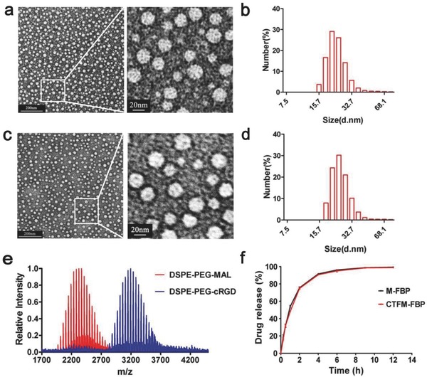
Figure 2.

Characterizations of nanomicelles. a) Transmission electron microscopy (TEM) images of M‐FBP. b) Dynamic laser scanning (DLS) measurement of M‐FBP. c) TEM images of CTFM‐FBP. d) DLS measurement of CTFM‐FBP. e) MALDI–TOF–MS analysis of conjugation of the cRGD peptide with DSPE‐PEG2000‐MAL. f) Time course of FBP release from M‐FBP and CTFM‐FBP nanomicelles in artificial tears over 12 h.
