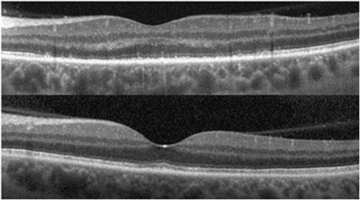
Figure 4.
Spectral-domain optical coherence tomography images of acute syphilitic posterior placoid chorioretinitis (top, visual acuity 6/18), demonstrating typical hyper-reflective nodular retinal pigment epithelial thickening and disruption of outer segment. Three months after treatment (bottom) the scan is anatomically normal (visual acuity 6/5).

