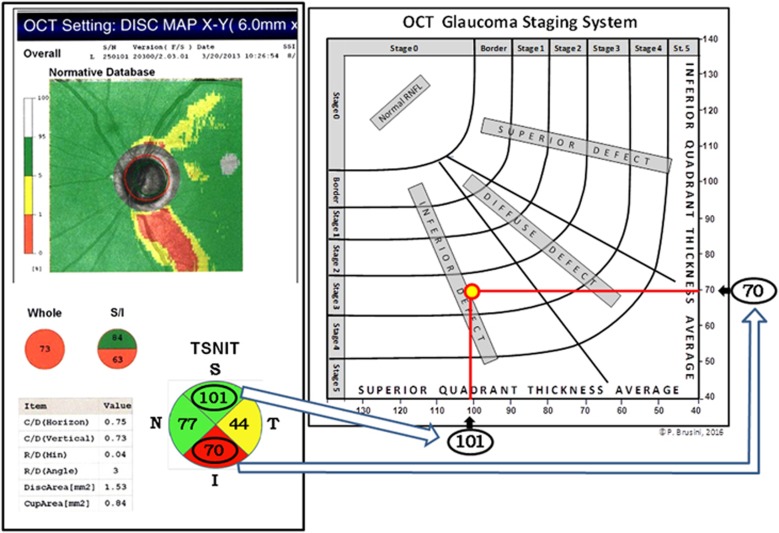
Figure 1.
Graphical representation of the OCT Glaucoma Staging System in a 61-year-old patient with POAG in the left eye. The intersection of the superior and inferior quadrant RNFL thickness values (arrows), expressed in microns, defines both the stage and the prevalent localization of the retinal nerve fiber defect.

