Figure 4.
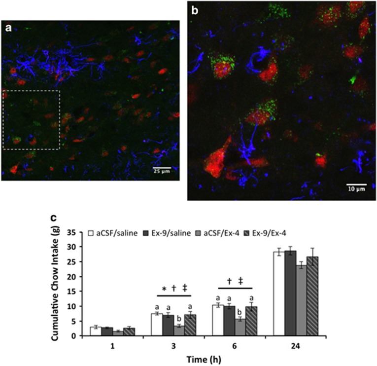
Systemically delivered GLP-1R agonists access the LDTg. To determine if peripherally administered Ex-4 penetrates the LDTg, we injected fluorescently tagged Ex-4 (FLEX, 3 μg/kg, IP), perfused the rats (n=5) 3 h later, and processed the LDTg to visualize neurons, astrocytes, and FLEX. Peripherally administered FLEX (green) is juxtaposed with neurons (red) but minimally with astrocytes (blue) within the LDTg (a,b). × 63 image in (a), and × 3 optical zoom of × 63 in (b). Dotted rectangle in (a) indicates field of view in (b) and in Movie 1. To determine if GLP-1R blockade in the LDTg attenuates the hypophagic effects of peripheral Ex-4, the competitive GLP-1R antagonist Ex-9 was unilaterally injected in the LDTg (n=9) at a dose subthreshold for an effect on feeding (10 μg; vehicle, 100 nl aCSF) approximately 1 h prior to the onset of the dark cycle. Fifteen minutes prior to the onset of the dark cycle, rats were injected systemically with Ex-4 (0 (saline), 3 μg/kg). Ex-4 significantly suppresses food intake at 3 h (p<0.05) and approaches significance at 6 h post-injection (p=0.06), and pre-treatment with Ex-9 reverses this intake suppression (c). A schematic map of accurate cannula placements for (c) can be found in Supplementary Figure S6B. * indicates significant main effect of Ex-4 (p<0.05). † indicates a significant main effect of Ex-9 (p<0.05). ‡ indicates a significant interaction between Ex-4 and Ex-9 (p<0.05). Different letters are significantly different from each other (p<0.05) according to post hoc tests. aCSF, artificial cerebrospinal fluid; GLP-1R, glucagon-like peptide-1 receptor; IP, intraperitoneal; LDTg, lateral dorsal tegmental nucleus.

