
Figure EV1. RAB7 localizes to mitochondria and the ER .
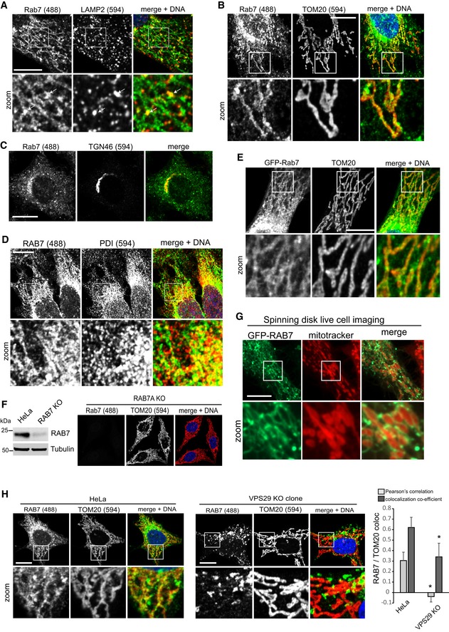
All images show formaldehyde‐fixed HeLa cells.
- HeLa cells stained for endogenous RAB7a (green) and endogenous LAMP2 (red). White arrows indicate sites of vesicular RAB7 that appears to be budding from a network of RAB7.
- Immunofluorescent co‐staining of endogenous RAB7a (green) and the mitochondria marker TOM20 (red).
- Co‐staining of endogenous RAB7 (green) and the trans‐Golgi network marker TGN46 (red).
- Co‐staining of endogenous RAB7 (green) and the ER marker protein disulfide isomerase (PDI, red).
- Co‐staining of lentivirally expressed GFP‐RAB7a and endogenous TOM20 (red).
- Co‐staining of endogenous RAB7 (green) and TOM20 (red) in cells treated with Cas9 and a mixture of three gRNAs targeting the RAB7a locus. The Western blot of this cell population shows an almost complete loss of RAB7 in the treated cells compared to parental HeLa cells.
- GFP‐RAB7 was expressed in RAB7a KO HeLa cells and imaged in live cells using a spinning disk confocal microscope. Mitochondria were visualized with MitoTracker Red.
- Parental HeLa cells and clonal VPS29 KO cells were co‐stained for endogenous RAB7a (green) and endogenous TOM20 (red), and co‐localization was analyzed across two independent experiments.