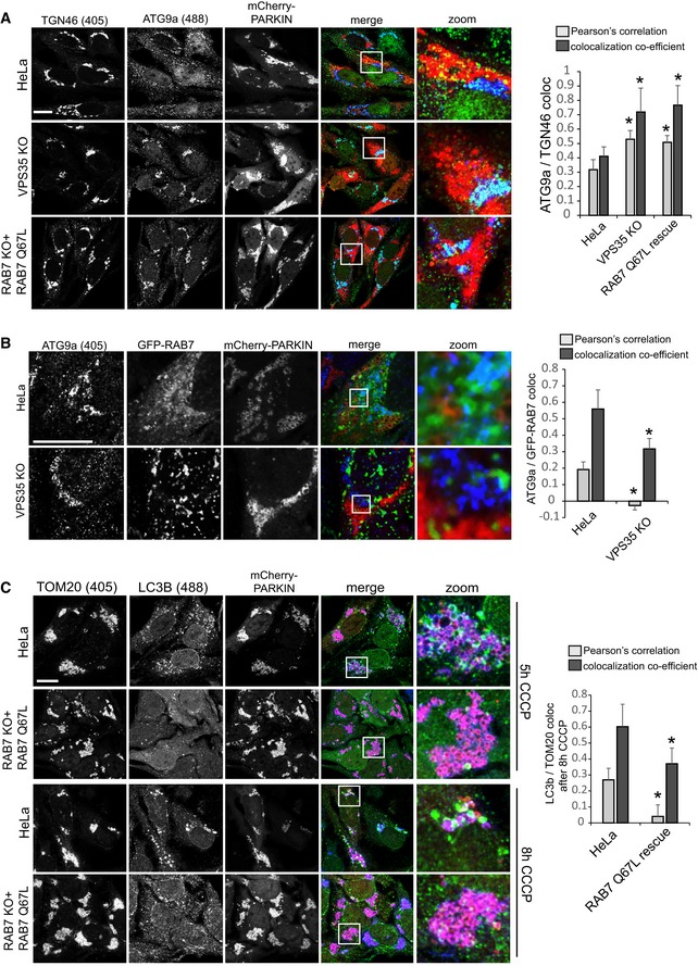
mCherry‐Parkin‐expressing parental HeLa cells, VPS35 KO cells, and RAB7 KO cells transduced with untagged RAB7‐Q67L were treated with CCCP for 5 h, fixed in PFA, and co‐stained for endogenous ATG9a (green) and the TGN marker TGN46 (blue). Co‐localization between ATG9a and TGN46 was analyzed across 14 images acquired in two independent experiments.
Parental HeLa cells and VPS35 KO cells were transduced with GFP‐RAB7 and mCherry‐Parkin and treated with CCCP for 5 h. The cells were then fixed in PFA and stained for endogenous ATG9a (blue). Co‐localization between GFP‐RAB7 and ATG9a was quantified across 14 images acquired in two independent experiments.
mCherry‐Parkin‐expressing parental HeLa cells and RAB7 KO cells transduced with untagged RAB7‐Q67L were treated with CCCP for 5 h (upper panel) and 8 h (lower panel), fixed in methanol, and stained for endogenous TOM20 (blue) and endogenous LC3b (green). Co‐localization between LC3b and TOM20 was quantified across 14 images acquired in two independent experiments.
Data information: All scale bars = 10 μm, all error bars = SD, and *
P < 0.05 in a
t‐test of the respective condition compared to the control cells.