Figure 1.

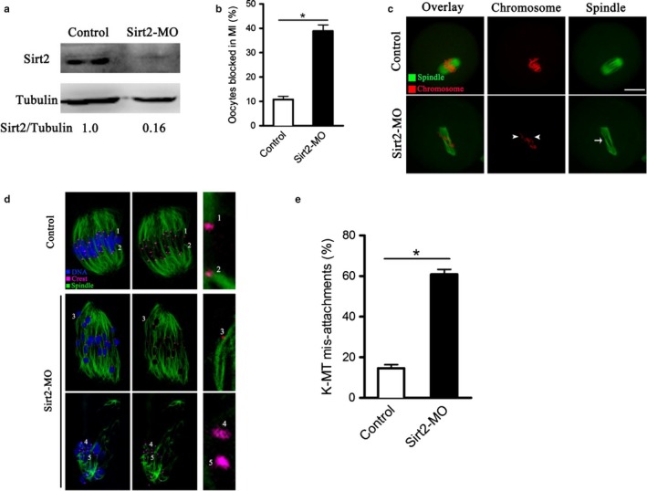
Effects of Sirt2 knockdown on maturational progression and meiotic structure in oocytes. Fully grown oocytes were injected with Sirt2‐MO and then cultured in vitro to evaluate the maturational progression and meiotic apparatus. (a) Western blotting showing the reduced expression of Sirt2 after MO injection. (b) Percentage of oocytes blocked in metaphase after Sirt2‐MO injection (n = 80 for control; n = 96 for Sirt2‐MO). (c) Control and Sirt2‐MO oocytes were stained with α‐tubulin antibody to visualize the spindle (green) and counterstained with PI to visualize chromosomes (red). Control metaphase oocytes present a typical barrel‐shape spindle and well‐aligned chromosomes. Spindle defects (arrow) and chromosome misalignment (arrowheads) were frequently observed in Sirt2‐MO oocytes. Representative confocal sections are shown. Scale bar, 25 μm. (d) Control and Sirt2‐MO oocytes at metaphase stage were labeled with α‐tubulin antibody to visualize spindle (green), CREST to detect kinetochore (purple), and co‐stained with Hoechst 33342 for chromosomes (blue). Representative confocal sections show the amphitelic attachment (chromosomes 1 and 2), and merotelic/lateral attachment (chromosome 3), and loss attachment (chromosomes 4 and 5) in Sirt2‐MO oocytes. (e) Quantification of control (n = 20) and Sirt2‐MO (n = 28) oocytes with K‐MT mis‐attachments. The graph shows the mean ± SD of results obtained in three independent experiments. *p < .05 vs. controls
