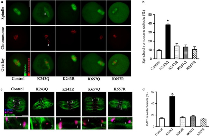
Figure 3.

Effects of BubR1 acetylation on meiotic apparatus and kinetochore–microtubule attachment in mouse oocytes. (a) Control and BubR1 mutant‐injected oocytes were stained with α‐tubulin antibody to visualize spindle (green) and counterstained with PI to visualize chromosomes (red). Representative confocal sections are shown. (b) Quantification of control and BubR1 mutant‐injected oocytes with spindle/chromosome defects. Data are expressed as mean percentage ± SD from three independent experiments in which at least 100 oocytes were analyzed. (c) Control and BubR1 mutant‐injected oocytes at metaphase stage were labeled with α‐tubulin antibody to visualize spindle (green), CREST to detect kinetochore (purple), and co‐stained with Hoechst 33342 for chromosomes (blue). Representative confocal sections are shown. BubR1‐K243Q indicates the loss attachment between kinetochore and microtubule. (d) Quantitative analysis of K‐MT mis‐attachments in control and BubR1 mutant‐injected oocytes. Data are expressed as mean percentage ± SD from two independent experiments in which at least 20 oocytes were analyzed. *p < .05 vs. controls
