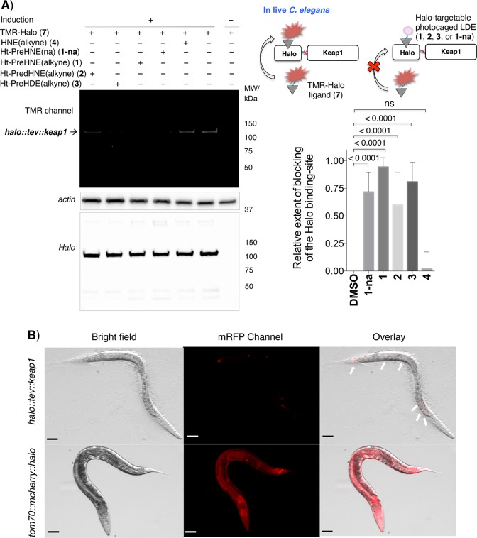
Figure 2.
(A) Photocaged precursors selectively bind to functional HaloTag expressed in live worms. The schematic at the top right shows the blocking experiment (readout A, Figure 1). See the Supporting Information for the detailed procedure. The left panel shows representative data analyzed by in-gel fluorescence (top) and Western blot (bottom) using anti-actin and anti-Halo antibodies, respectively, that confirm protein loading and inducible halo::tev::keap1 expression (halo::tev::keap1 full construct molecular weight of ∼105 kDa). See also Figure S2 for additional replicates. In the bottom right panel, the ratios of the fluorescence signal to the anti-Halo Western blot signal were normalized to dimethyl sulfoxide within each independent gel before averaging across multiple replicates. Errors designate the standard deviation (n = 8 independent biological replicates). (B) Fluorescence images of the heat shock-induced live worms further confirm transgene expression. Fluorescence of tom70-MLS-localized mcherry::halo is visible throughout the worm (bottom row). While halo::tev::keap1 does not itself feature a fluorescent marker, it is co-expressed with a constitutive dominant marker (mec7p::mrfp), which displays fluorescence localized to touch-receptor neurons (top row, white arrows). Scale bars are 50 μm.

