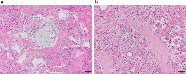
Fig. 2.

a Transbronchial lung biopsy showed proliferative-phase diffuse alveolar damage with a glassy eosinophilic substance in the alveolar spaces and interstitial fibrosis associated with type 2 pneumocyte hyperplasia. b Autopsy lung section showed fibrotic-phase diffuse alveolar damage with diffuse fibrotic change and type 2 pneumocyte hyperplasia, with lymphocyte infiltration into alveolar septa
