Fig. 2.

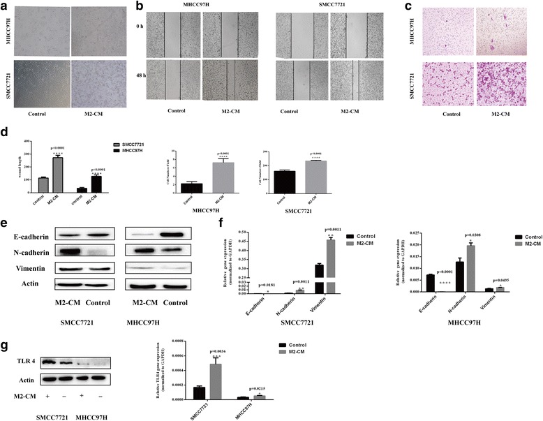
M2-CM increased the malignant properties of HCC cells and induced TLR4 activation. a M2-CM increased the number of HCC cells with the fibroblast-like morphology (magnification, × 100). b Wound-healing assay. Wound closure was delayed in M2-CM-treated MHCC97H and SMMC7721 cells compared with in the control group at 48 h (magnification, × 50). c Transwell migration assays. The number of cells passing through the upper chamber was counted in four fields (magnification, × 100). d Analysis of the results of the wound-healing assay and transwell migration assay. e–f M2-CM promoted EMT in HCC cells. The expression of EMT markers E-cadherin, N-cadherin, and vimentin in M2-CM-stimulated HCC cells, and the control group was analyzed using western blots and RT-PCR. g M2-CM induced TLR4 activation in HCC cells. The expression of TLR4 on HCC cells in M2-CM and control cells was detected using western blots and RT-PCR. Date are shown as the means ± SD (*P < 0.05, **P < 0.01, ***P < 0.001, ****P < 0.0001). The data represent at least three independent experiments
