
Fig. 1.
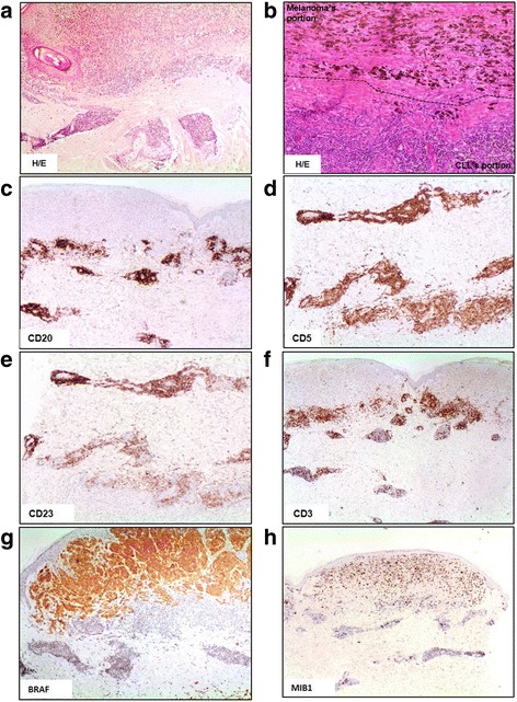
Immunohistochemical characterization of skin lesion. a Haematoxylin/eosin staining, ×4. b Haematoxylin/eosin staining, ×10, the dotted black line defines the border area between CLL and MM tumors. c, d and e Immunohistochemical positivity for CD20, CD5 and CD23 antigens. f Immunohistochemical negativity for CD3. g Presence of mutant BRAF only in MM lesion. h MIB1 expression to evaluate the neoplastic proliferation