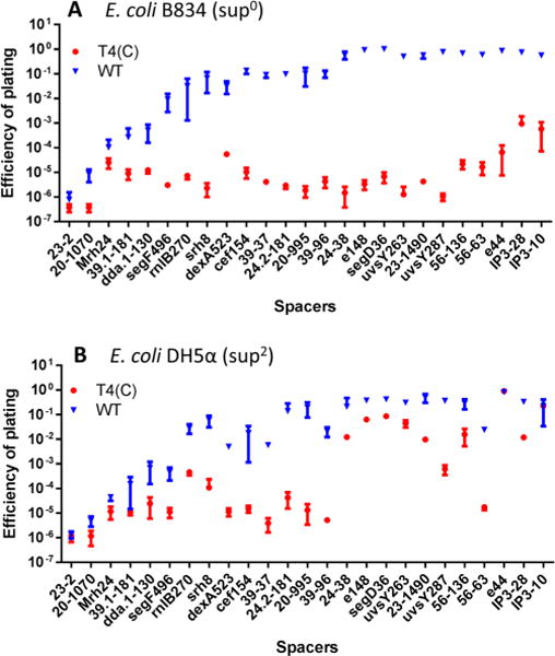
Figure 2.

Restriction of phage T4 infection by CRISPR-Cas. The spacer-containing E. coli cells were infected with WT T4 or T4(C) mutant as per the basic scheme shown in Figure 1. See Materials and Methods for more details. The restriction of phage T4 infection was determined by the efficiency of plating (EOP), as determined by plaque assay. EOP was calculated by dividing the number of pfu produced on the spacer containing E. coli by the number of input pfu. EOP of modified (WT, blue symbols) or unmodified mutant (T4(C), red symbols) phages was determined on E. coli strains B834 (sup0) (A) or DH5α (sup2) (B). The labels on the X-axis denote the spacer. The sequences of the spacers are listed in Table 1. The experiments were done in triplicate.
