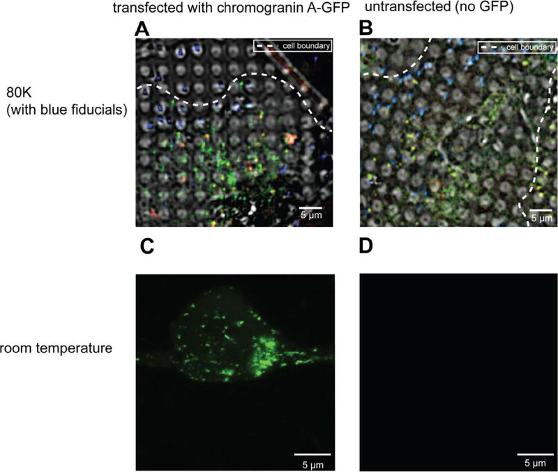
Fig. 1.
INS-1E cells exhibit strong autofluorescence at 80 K. (A, B) Deconvolved cryo-LM images (composite of bright field and epifluorescence in FITC, mCherry and DAPI channels) of INS-1E cells transfected with CgA-GFP (A) or untransfected (B). (C, D) Room-temperature light microscopy images of epifluorescence in FITC channel of INS-1E cells transfected with CgA-GFP (C) or untransfected (D).

