
Fig. 1.
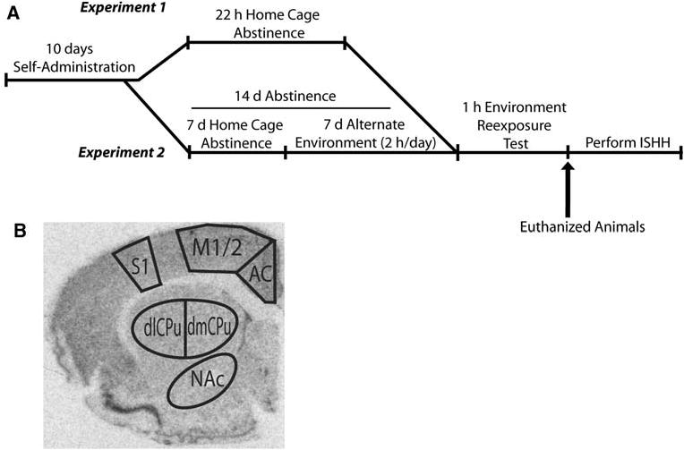
a Schematic representing the experimental design for experiment 1 and 2 (adapted from Hearing et al. 2008). SA and CA rats were not included in the design of experiment 1 because exposure to an alternate environment on day 1 of abstinence would have constituted a novel experience. In experiment 2, all rats were habituated to the alternate environment before the test day. b Audioradiographic image from tissue labeled with 35S-dATP-labeled zif/268 oligonucleotide illustrating brain regions analyzed. Regions of interest selected for measurement according to Paxinos and Watson 2007. AC anterior cingulate, dlCPu dorsolateral striatum, dmCPu dorsomedial striatum, M1/2 motor1/2 cortex, NAc nucleus accumbens core, S1FL sensory 1 forelimb cortex, SN saline alternate environment, CA cocaine alternate environment