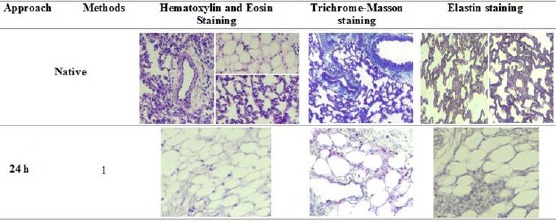
Figure 2.

Hematoxylin and Eosin Staining showed structures and nuclei present in native and decellularized lungs. Trichrome-Masson staining showed collagen levels in native and decellularized lungs (Blue colour). Elastin staining showed elastin levels in native and decellularized lungs (Blue colour to black colour). Representative images for all conditions are shown (100 × magnification)
