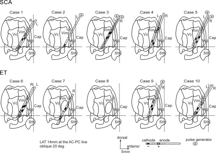
Figure 3.

The best locations of the clinically defined therapeutic DBS contacts and the coagulation lesions presented on the parasagittal section with an oblique dorsolateral to medioventral angle of approximately 20 degrees (adapted from the Shaltenbrand and Bailey Atlas18). The common finding was that at least one cathodal contact was located in the lower and posterior quadrant in the sagittal section of the Vim in most patients in SCA and ET. The lateral distance of the best contacts was 12–16 mm from the midline. The coagulation areas were located in the central area of the Vim in the thalamotomy cases. The horizontal broken lines are the anterior commissure–posterior commissure lines (AC‐PC lines), and the vertical broken lines are the mid AC‐PC lines. Cap, capsule; Sth, subthalamic nucleus; Vc, nucleus ventrocaudalis; Vim, nucleus ventrointermedius; Vop, nucleus ventrooralis posterior.
