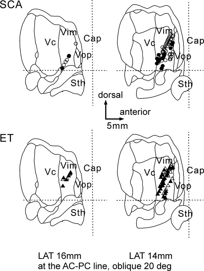
Figure 4.

Location of neuronal recording. The positive sensory response was shown by filled circles and triangles. The recording areas were apparently no difference between SCA and ET. The horizontal broken lines are the AC‐PC lines, and the vertical broken lines are the mid AC‐PC lines. Cap, capsule; Sth, subthalamic nucleus; Vc, nucleus ventrocaudalis; Vim, nucleus ventrointermedius; Vop, nucleus ventrooralis posterior.
