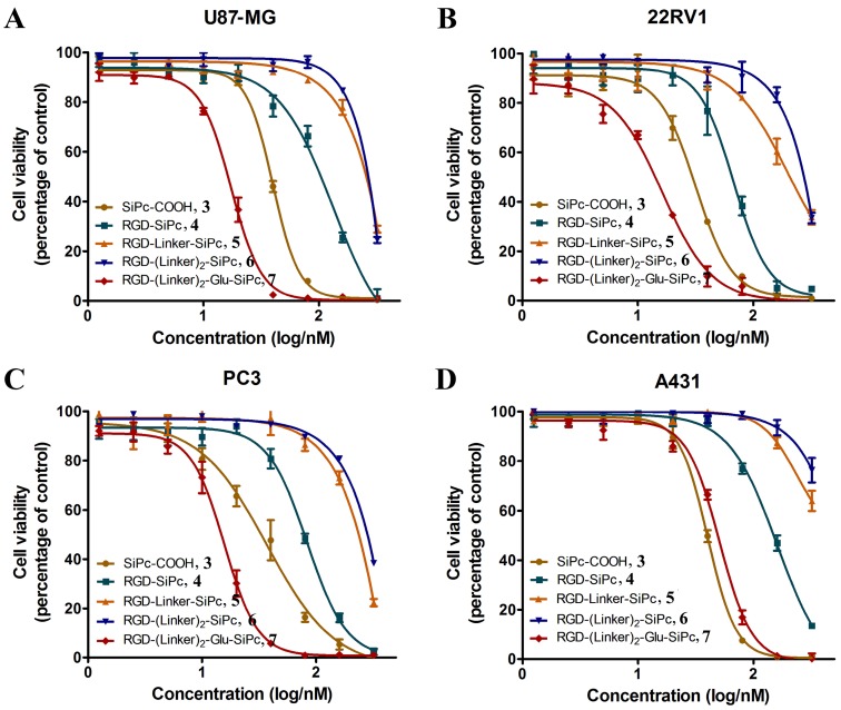
Figure 4.
In vitro concentration-dependent photocytotoxicity of SiPc-COOH 3 (circles, dark yellow), RGD-SiPc 4 (squares, olive), RGD-Linker-SiPc 5 (upward triangles, orange), RGD-(Linker)2-SiPc 6 (downward triangles, blue) and RGD-(Linker)2-Glu-SiPc 7 (diamonds, red) on receptor-positive U87-MG (A), 22RV1 (B), and PC3 (C) cells and receptor-negative A431 cells (D) (n = 3, mean ± SEM), as determined by MTT assay. The cells were treated with various concentrations of the conjugates for 4 h before illumination with light (40 mW/cm2, 15 min), and cell viability was determined by MTT assay.

