
Figure 6.
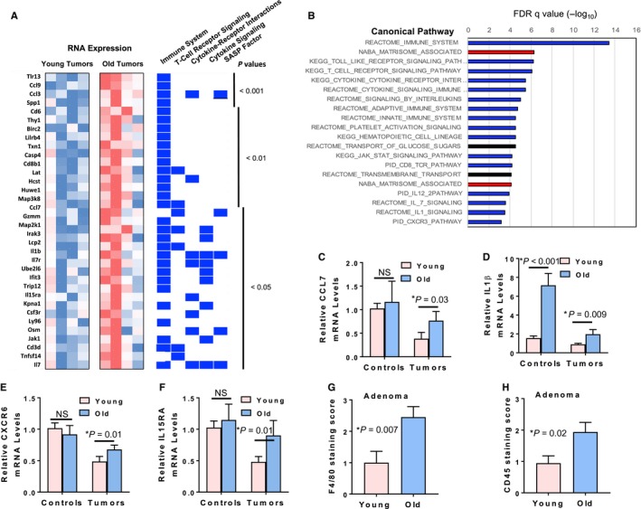
Lung adenomas from old Kras G12D mice show upregulation of genes affiliated with immune cells and inflammatory pathways. (A) Microarray results show enhanced expression of multiple inflammatory pathway genes in lung tumors from old Kras G12D mice. Representative upregulated genes in old Kras G12D tumors with P value < 0.05 were compared for relative expression in young and old tumors as shown in the heat map. Each gene was also assessed for association (indicated by blue rectangles) with immune cell and cytokine pathways using GSEA. The last category, ‘SASP Factor’, indicates those genes associated in the literature with the senescence‐associated secretory phenotype. (B) Genes overexpressed in old Kras G12D tumors show enrichment for immune cell and inflammatory pathways by GSEA. The top 20 most significantly enriched canonical pathways are shown based on GSEA of the 176 significantly upregulated genes in old Kras G12D tumors relative to young Kras G12D tumors. Relative P values (−log10) are indicated above the graph. Blue bars indicate immune cell/cytokine/inflammatory pathways, and red bars indicate extracellular matrix‐associated pathways. (C–F) qPCR validation shows significant upregulation of inflammation‐related genes Ccl7 (C), IL‐1 β (D), Cxcr6 (E) and IL‐15ra (F) in lung tumors from old Kras G12D mice (n = 5 for young and old mice). For control samples (n = 5 for young and old mice), only IL‐1‐β showed significant upregulation in old mice (D). (G–H) Immune cells cluster in close proximity to the lung tumors derived from old mice. Lung adenomas from young and old Kras G12D mice stained for macrophages (F4/80 antibody, G) and leukocytes (CD45 antibody, H) were visually assessed and scored on a scale of 0–4 by a pathologist based on the estimate of immune cells present within the tumor or within 100 μm of the tumor boundary. Twenty lung tumors from six old mice and 14 lung tumors from five young mice were examined for F4/80 staining. Twenty‐four lung tumors from five old mice and 18 lung tumors from six young mice were examined for CD45 staining.