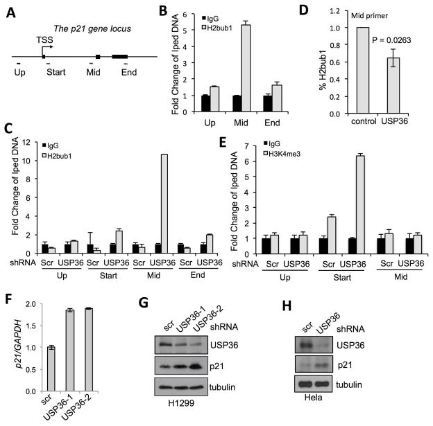
Figure 3. USP36 deubiquitinates H2Bub1 within the p21 gene.
(A). A schematic diagram of the p21 gene locus. Filled boxes indicate exons. Primers for amplifying different regions relative to transcriptional start site were indicated. (B). H2Bub1 levels are enriched within the p21 gene body. 293 cells were subjected to ChIP using control IgG or anti-H2Bub1, followed by qPCR detection using indicated primers. (C). Knockdown of USP36 increases H2Bub1 in the p21 gene body. H1299 cells infected with scrambled (scr) or USP36 shRNA were assayed by ChIP using anti-H2Bub1 or IgG, followed by qPCR. (D). Overexpression of USP36 reduces the levels of H2Bub1 in the p21 gene body. H1299 cells transfected with control or Flag-USP36 followed by ChIP using anti-H2Bub1 and qPCR detection of the p21 gene body. (E). Knockdown of USP36 increases H3K4me3 near the transcription start sites of the p21 gene. H1299 cells infected with scr or USP36 shRNA were assayed by ChIP using anti-H3K4me3 or control IgG, followed by qPCR. (F) (G). Knockdown of USP36 increases p21 expression in cells. H1299 cells were infected with scr or USP36 shRNA-encoding lentiviruses, followed by detection of p21 mRNA by qPCR (F) and p21 protein by IB (G). (H) HeLa cells infected with scr or USP36 shRNA were assayed by IB.

