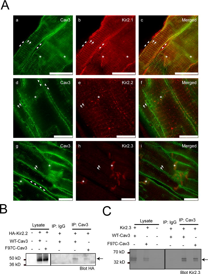
Figure 1.

A) Kir2.1, Kir2.2 and Kir2.3 coimmunolocalize with Cav3 at specific subcellular domains in human ventricle. STED images of human left ventricle tissue stained for Kir2.1 (b, red), Kir2.2 (e, red), Kir2.3 (h, red) and Cav3 (a, d and g in green). The merged panels are c, f and i. Arrows represent T-tubular pattern of staining. Arrow heads identify the lateral membrane. Asterix identify intercalated disk. Scale bar = 10 μm. B) and C) Kir2.2 and Kir2.3 co-imunoprecipitate with Cav3. HA-Kir2.2 (B) and Kir2.3 (C) co-immunoprecipate with WT-Cav3 and F97C-Cav3 in HEK293 cells as shown by arrows.
