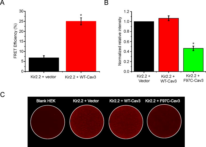
Figure 3. F97C-Cav3 significantly reduces Kir2.2 cell surface expression in HEK293 Cells.

A) The bar graph describes the summary data from multiple FRET experiments between Kir2.2 (GFP tagged) and Cav3 (mCherry tagged), showing that the average FRET efficiency is 25+/−1.76% (n=18) as compared with 6.86+/−1.05% (n=19) in control (Kir2.2 (GFP tagged) + vector (mCherry)) p<0.001. (* indicates p<0.001) B) The bar graph displays quantitative results of eight independent experiments p<0.001. (* indicates p<0.001) C) Representative on-cell Western blot analysis illustrating intensity levels of extracellular HA-tagged Kir2.2 surface expression in wells co-transfected with HA-Kir2.2. Panel (a) shows vector only, Kir2.2 alone panel (b), Kir2.2 with WT-Cav3 (c), and F97C-Cav3 (d). a shows untreated cells stained with both primary and secondary antibody.
