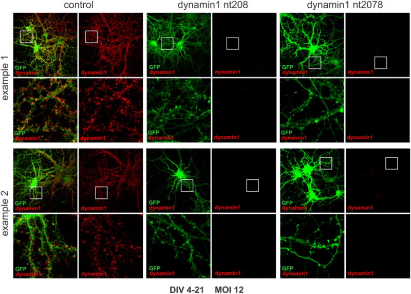
Figure 2. Immunofluorescence analysis of dynamin 1 expression in control and dynamin 1 knockdown neurons.

Primary rat hippocampal neurons were transduced on DIV 4 with control or dynamin 1 knockdown shRNAmiR viruses as indicated using an MOI of 12 and processed for immunofluorescence on DIV 21. Two examples for each condition are shown. GFP is the fluorescent marker protein expressed as part of the shRNAmiR cassette and dynamin 1 was detected using an antibody directed against endogenous dynamin 1. Dynamin 1 is readily detected in neurons transduced with the control virus but is virtually undetectable in neurons transduced with the knockdown viruses. The areas in the white boxes seen in the lower magnification images is shown at higher magnification below each image.
