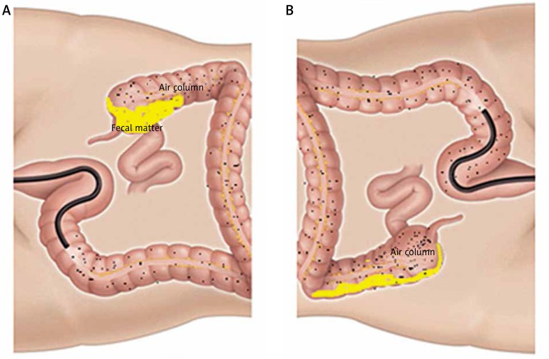
Figure 2.
This figure implicate the possible mechanism of improved caecal visualisation with changing the position from left lateral (A) to right lateral (B). The formed faecal matter in the caput of caecum, which is the dependant part on the left lateral position of the patient, decreases the caecal visualisation. Changing the position to supine or right lateral moves the air column at the caecal base with the gravitational forces in action making the caput a non-dependant area, which results in the movement of the formed faecal matter from the caput of caecum

