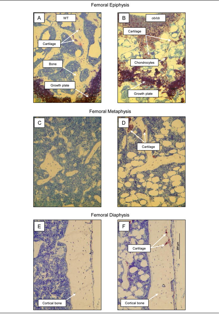
Figure 2.

Photomicrographs of the distal femur epiphysis, metaphysis, and diaphysis in representative 4-month-old male WT mice (A, C, E) and 4-month-old male leptin-deficient ob/ob mice (B, D, F). Whereas cartilage matrix was rare in the epiphysis in WT mice (A), cartilage matrix and chondrocytes were readily visible in the epiphysis of ob/ob mice (B). Leptin-deficient ob/ob mice also exhibited a highly disorganized growth plate architecture, characterized by irregular margins and width (B). Cartilage was rare in the metaphysis and diaphysis of WT mice (C and E, respectively), but present at both sites in ob/ob mice (D and F).
