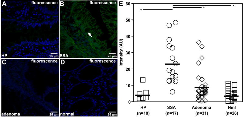
Fig. 6. Ex vivo peptide binding to human colon specimens.
A–D) On confocal microscopy, KCC*-FITC (green) binds brightly to surface (arrow) of colonocytes from sessile serrated adenoma (SSA), while minimal signal is seen for hyperplasia (HP), adenoma, and normal colonic mucosa. E) The mean (±std) fluorescence intensities for HP (n = 10), SSA (n = 17), adenoma (n = 31), and normal (n = 26) were found to be 4.04±1.12, 22.93±3.07, 8.81±1.63, and, 3.56±0.64 respectively. The mean fluorescence intensity for SSA was found to be significantly higher than that for HP, adenoma, and normal, *P=<0.01, by unpaired t-test.

