
Figure 33.
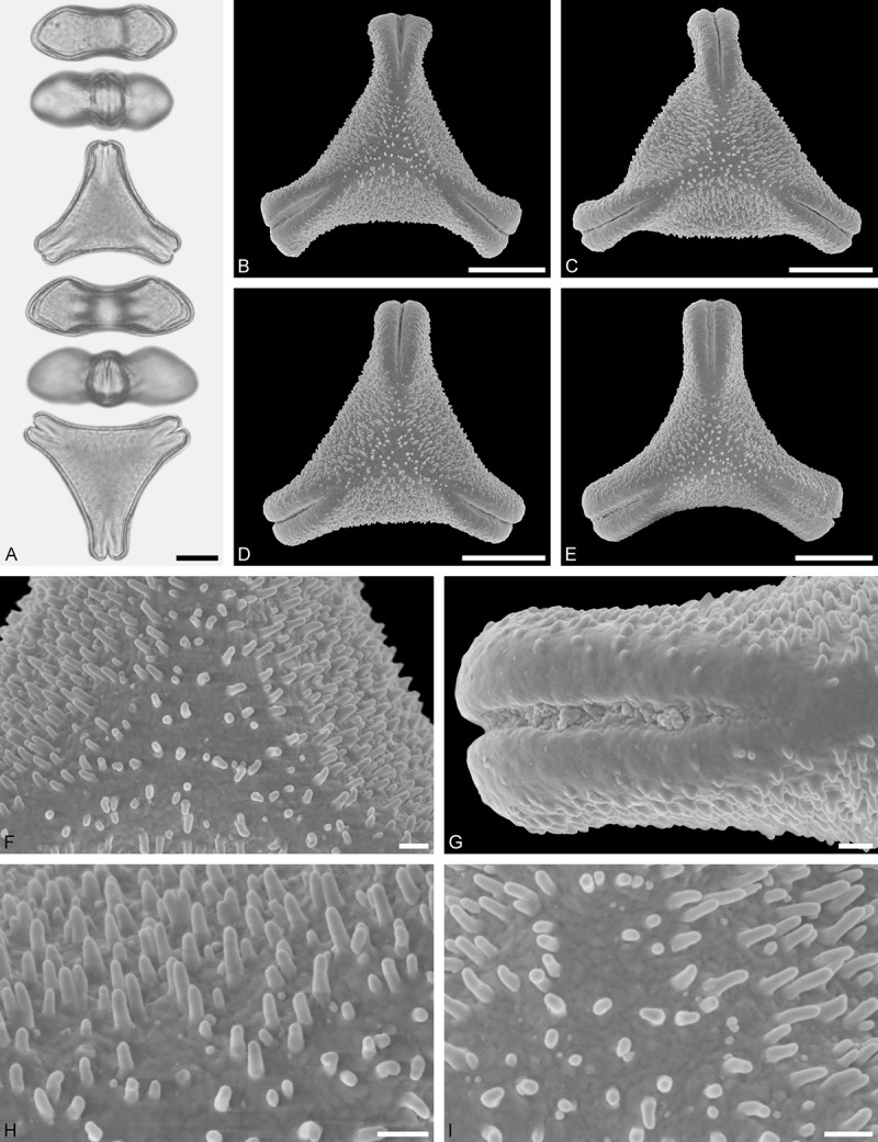
LM (A) and SEM (B–I) micrographs of Psittacanthus rhynchanthus (WU 020859). A. Two pollen grains in equatorial and polar view. B–E. Pollen grains in polar view. F. Close-up of central polar area. G. Close-up of apex. H. Close-up of mesocolpium. I. Close-up of central polar area. Scale bars – 10 µm (A–E), 1 µm (F–I).