
Figure 60.
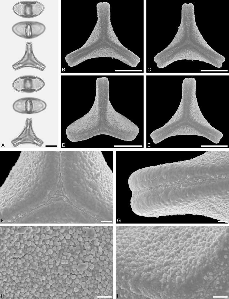
LM (A) and SEM (B–I) micrographs of Plicosepalus curviflorus (WU: from Kenya, coll. J. B. Gillett, s.n.). A. Two pollen grains in equatorial and polar view. B–E. Pollen grains in polar view. F. Close-up of central polar area. G. Close-up of apex. H, I. Close-ups of mesocolpium. Scale bars – 10 µm (A–E), 1 µm (F–I).