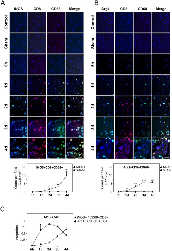
Fig 4. CD8-expressing cells in post-stroke brain.
(A–B) Upper panel shows representative triple-labeled immunostaining of CD8+CD68+ cells expressing iNOS or Arg1 in the perilesional areas of the ischemic hemisphere at 6 h, and at days 1, 2, 3 and 4 after stroke. Lower panel shows quantification data. (C) Proportion of CD8+CD68+ cells expressing either Arg1 or iNOS at 4 d after stroke. Data in A–C is presented as average ± SEM. Scale bars represent 32 μm. * P < 0.05; ** P < 0.01; *** P < 0.001.

