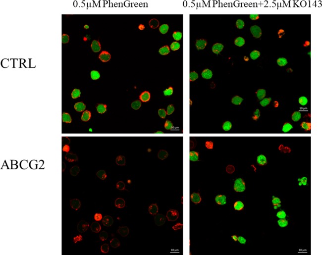
Fig 3. Fluorescent PG accumulation in human PLB cells, examined by confocal microscopy.

Effects of ABCG2 protein expression and the specific inhibition of ABCG2 function by Ko143. Cellular fluorescence was observed by confocal microscopy. PG fluorescence (green) was examined after 30 minutes of the addition of 0.5μM PGD to the medium, either in the absence or presence of the ABCG2 inhibitor KO143 (2.5μM). The cells were pre-labeled with fluorescent anti-WGA (red) to indicate the plasma membranes.
